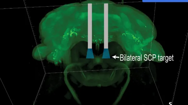
The brain’s internal clock may help determine when seizures occur
Research



New research led by Dr. Vishnu Anand Cuddapah, Principal Investigator at the Jan and Dan Duncan Neurological Research Institute at Texas Children’s Hospital and Assistant Professor of Pediatrics, Neurology at Baylor College of Medicine and published in Nature Communications, has identified brain cells that determine when seizures happen. The findings reveal that neurons responsible for regulating the body’s internal clock can switch between signals that increase seizure risk during the day and signals that suppress it at night.
More than 80% of people with epilepsy exhibit daily patterns in seizure risk, although the time when seizures are most likely to occur varies among individuals. Researchers have long suspected that circadian rhythms, the approximately 24-hour biological cycles that regulate sleep, metabolism, hormones and other processes, could play an important role. Separating the effects of the circadian clock from sleep and other daily changes, however, has been difficult.
To investigate the underlying mechanisms, the researchers turned to the fruit fly. Fruit flies also have circadian systems and provide scientists with powerful genetic tools for studying the activity of individual groups of neurons. Their brains contain approximately 240 clock neurons that help coordinate daily behavioral rhythms.
The team developed a system that recorded fruit flies for 96 hours and automatically identified seizure activity. Researchers found that, much like seizures in people, seizures in the fruit flies followed a distinct daily pattern and occurred more frequently during the daytime.
When the researchers disrupted a core gene involved in the fruit flies’ molecular circadian clock, that daily rhythm disappeared. Seizures still occurred, but their timing became randomly distributed throughout the day. Importantly, the researchers found that these seizure rhythms were independent of the amount of sleep the fruit flies received, suggesting that the internal biological clock itself was influencing seizure susceptibility.
The team then narrowed the effect to specific groups of clock neurons. They discovered that ventrolateral neurons, including cells known as “morning cells,” and dorsolateral neurons, or “evening cells,” help regulate the daily rhythm in seizure risk.
During the day, morning cells release the molecules pigment-dispersing factor (PDF) and short neuropeptide F (sNPF). These signals increase activity across the brain and make seizures more likely to occur. At night, however, the same morning cells appear to switch their communication strategy, using the neurotransmitter glycine to reduce seizure susceptibility, likely through their effects on evening cells.
The result is a biological switch in which the brain’s clock circuitry can promote seizure susceptibility at one time of day and suppress it at another. The investigators further tested the role of these neurons using optogenetics, a technique that allows scientists to control selected neurons with light. Inhibiting clock neurons reduced seizure risk, providing additional evidence that these cells actively regulate seizure susceptibility rather than merely reflecting other changes occurring throughout the day.
The findings provide a new biological explanation for what doctors have observed in epilepsy patients. This research on fruit flies translates to the human brain as components of circadian biology are conserved across species, making the discovery an important starting point for additional studies. By uncovering a direct connection between the brain’s internal clock and seizure susceptibility, the research contributes to more precise approaches to epilepsy treatment.
###
Other contributors to this work include Cynthia T. Hsu, Chitrakshi Pant, Camilo Guevara, Jianing Yang, Samantha Killiany, Hrishit M. Shah, Mary E. Putt, and Amita Sehgal. The authors are affiliated with one or more of the following institutions: Duncan NRI, Texas Children’s Hospital, Baylor College of Medicine, the University of Pennsylvania, and the Children’s Hospital of Philadelphia.
This work was supported in part by National Institutes of Health (NIH) grant K08NS131602, P50HD105354, R01NS048471, CURE Epilepsy Taking Flight Award, Howard Hughes Medical Institute, the McNair Medical Institute at the Robert and Janice McNair Foundation, the Gordon and Mary Cain Research Foundation Laboratories, and the Research Vision Scholars program at Texas Children’s Hospital.