Topics
Nine years ago, at their 20-week ultrasound check, Keri and Chad McCartney were thrilled to learn their fifth and final child would be a girl. But that excitement quickly turned to fear when the image on the screen caused the technician to fall silent.
The ultrasound revealed a grapefruit-sized mass, called a sacrococcygeal teratoma (SCT), growing out of the baby’s tailbone. It was about the same size as the baby. This type of very rare tumor, found in only 1 of 35,000 pregnancies, was drawing on her blood supply and would ultimately lead to heart failure.
“Our OB/GYN explained that our baby’s odds of survival were less than 10% because the tumor was so blood-filled,” recalls Keri. “We were completely heartbroken.”
The McCartney’s OB/GYN searched for help across the U.S. and Texas Children’s Fetal Center responded. A team of nearly 30 specialists met to evaluate the McCartney baby’s case and came to a unanimous decision.
Despite the fact that few operations of its kind had ever been performed or even been successful, fetal surgery offered the McCartney’s the potential for a better outcome.
“For the first time, we at least knew that good or bad the outcome, we were in a place where they would walk us through the whole thing,” says Chad. “If there was any place this could happen, Texas Children’s was the place.”
With Macie’s heart failing, the McCartney’s had one day to make a decision and they decided they couldn’t say no to their baby. The surgery was scheduled for the next day.
“It brought me such comfort during that time that as sick as she was, a 25-week baby, that they saw her life worth fighting for,” says Keri. “We talked to the doctors and knew that if things didn’t work out that we’d fought for her and done everything we could.”
On February 28, 2008, a surgical team led by Dr. Oluyinka Olutoye opened Keri’s womb, partially removed her 25-week-old daughter, and cut away most of the tumor in a single 4-hour procedure. Keri was given more than seven times the normal amount of anesthesia to prevent labor and protect the baby.
A critical test came when the tumor, which filled Olutoye’s hand, was removed. Would the heart respond positively? The answer was a resounding yes, and baby Macie continued to develop safe inside her mother’s womb.
Ten weeks after the surgery at 35 weeks, Macie Hope McCartney made her second appearance in the world, this time via C-section. Weighing in at 6 lbs, 1 oz., Macie was a healthy baby girl.
It was no coincidence that her parents gave her the middle name of Hope.
Macie was discharged from Texas Children’s one month later and joined her four siblings at home in Laredo. Besides the small scar on her tailbone, Macie shows no signs or indications that she endured such a life-threatening experience.
“If we didn’t tell people, no one would know,” says Chad. “There’s no evidence, apart from the scar, that lets you know she’s gone through anything of that nature. It’s just incredible.”
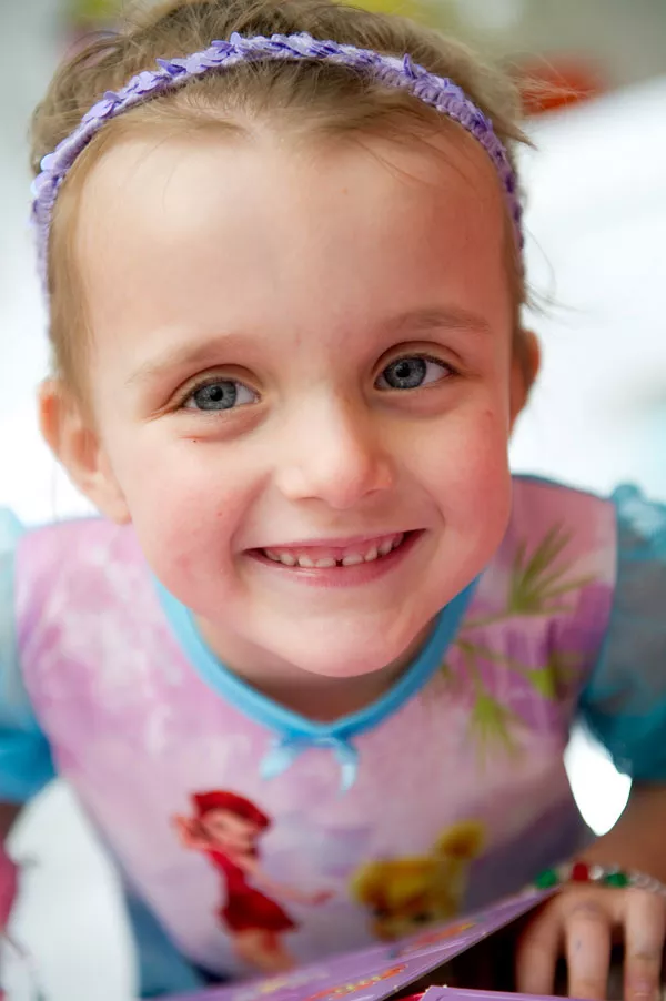
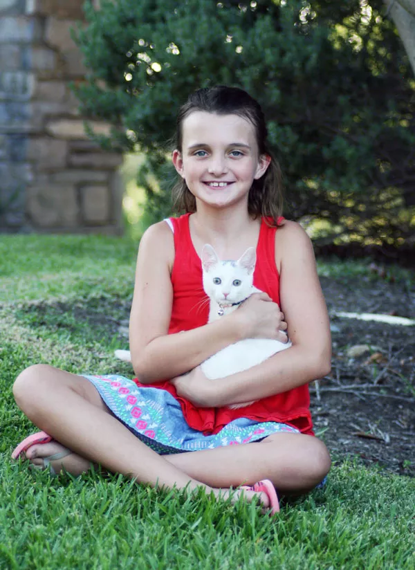
Today, Macie is a thriving, playful 9-year-old, and remains every bit the miracle girl she was before her birth.
“We turned to Texas Children’s because we wanted that hope that they would be able to help her,” recalls Keri. “We are humbled and thankful because we received a gift that not everyone is lucky enough to get.”


What is a sacrococcygeal teratoma?
A sacrococcygeal teratoma (SCT) is rare tumor located at the base of the coccyx, or tailbone. It is the most common tumor found in newborns though it only occurs in about one in 35,000 births. It is more common in female than male babies.
SCTs may grow very large, but are usually not malignant (cancerous). An SCT may be suspected if the mother’s blood work shows a high alpha fetoprotein or if a sonogram shows the uterus is larger than it should be. SCT is diagnosed with an ultrasound exam.
A sacrococcygeal teratoma is categorized by its location and severity:
- Type I tumors are outside the body and are attached to the tailbone.
- Type II tumors have both internal and external parts.
- Type III tumors can be seen from the outside, but most of the tumor is found in the fetus’ abdomen.
- Type IV tumors, the most severe type, exist completely inside the body at the tailbone level.
Less severe SCTs may be removed after birth. More severe SCTs may require fetal surgery in order to give the baby a chance to survive.


