Updates

Cloacal Malformation

Conditions

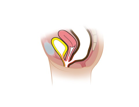
A cloacal malformation, or cloaca, is a rare condition that occurs in 1 in 25,000 girls. It is a severe form of anorectal malformation in which the rectum, vaginal, and urinary tract do not separate but merge into one channel, called a cloaca. The defect occurs during fetal development when the rectum, vaginal and urinary tracts fail to separate.


Symptoms

Symptoms may include the following:

  • The baby does not have a visible anus
  • An abnormal closing or absence of a vagina
  • A single opening in the baby’s pelvic area
  • Small external genitalia with an oversized clitoris

Diagnosis

  • A cloacal malformation may be detected during pregnancy with an ultrasound. If a cloaca is diagnosed during pregnancy, the fetus is monitored throughout the pregnancy with follow-up ultrasounds.
  • After birth, cloacal malformations may be discovered by physical examination or if the baby’s first bowel movement does not occur or passes out of the wrong opening. If the malformation is suspected by physical examination, the doctor may order diagnostic images, such as an ultrasound. Frequently, an exam under anesthesia is performed. This can give us important information about the anatomy that is difficult to obtain with imaging studies alone.

Treatment

Cloacal malformations require surgery. At Texas Children’s Hospital, a multidisciplinary team of doctors from pediatric surgery, urology, pediatric and adolescent gynecology, and gastroenterology work together to treat your child’s condition.

Colostomy

After birth, surgery will be performed to create an ostomy. In this operation, the intestine is connected to a surgical opening created in the abdomen (stoma) so that stool may pass into a bag outside the body. The ostomy is allows the child to pass stool until they can undergo their reconstructive surgery. The ostomy is usually reversed after reconstructive surgery or once the child can pass stool through their anus.

Vaginostomy

This is a procedure to provide drainage of the urinary tract through decompression of the gynecologic system where the urine tends to accumulate. This involves placing a tube into the vagina that is connected to a bag where the urine can drain. This is often done after birth at the time of colostomy formation. Some children drain urine freely and do not need this procedure. Other infants have a common channel that is easy to pass a catheter into, and can be managed with intermittent catheterization instead of a vaginostomy. It is imperative however to make sure the urine is being completely drained, because otherwise it can back up and cause irreversible damage to the kidneys. Our team will follow your child’s kidneys and bladder closely with ultrasounds to make sure the urine is emptying well with whatever method of drainage is used.

Exam under anesthesia

Around three to six months of age, we do an exam under anesthesia in the operating room with gynecology, urology and general surgery. This is where we learn all the details we need to know about the anatomy before planning reconstructive surgery. This involves placing a very small camera into the common channel, bladder, and vagina to visualize the anatomy and measure the length of the urethra and common channel. We also stimulate the anal sphincter muscle complex to determine its location and determine how strong that muscle is. This give us some information that helps us predict future bowel continence. These procedures are short, and you will go home the same day.

Reconstructive Surgery

Around six to twelve months of age, reconstructive surgery will be performed to separate the anus, urethra, and vagina. Once your child has healed from the reconstruction, a surgeon will reverse the colostomy to allow your child to achieve bowel movements through their anus.


Prognosis (or Long-Term Outcomes)

Our ultimate goal for your child is to be clean and dry of urine and stool, and to be able to function sexually later in her life. Even after surgery to correct the anatomy, some children with cloacal malformations will have issues with fecal or urinary incontinence.

Bowel control

Our bowel management team at Texas Children’s Hospital will help follow your child after surgery and through potty training into childhood to help with stooling. The quality of the sacrum, the presence or absence of any spinal cord anomalies such as tethered cord, and the length of the common channel are all key predictors of bowel control. Our bowel management team will work with you to help your child achieve continence.

Urinary Control

Our urology team will closely follow your child’s urinary continence at our multidisciplinary clinics. Some patients may need intermittent bladder catheterization to help empty their bladder and keep them clean and dry of urine in between catheterizations.

Gynecologic function

As your daughter approaches puberty, our gynecology team will identify any problems that may occur with menses. Before the patient becomes sexually active, our gynecologists will evaluate the vaginal opening to make sure it does not need any additional repairs or dilation. Our team will help counsel you and your daughter about sexual activity, fertility, and child bearing as she reaches the appropriate maturity.